2021 2nd Place Emage Winner: Scurvy
Case Presentation
A 2-year-old otherwise healthy female presented with new petechial rash and non-weightbearing on bilateral lower extremities in the context of one month of intermittent fevers and arthralgias.
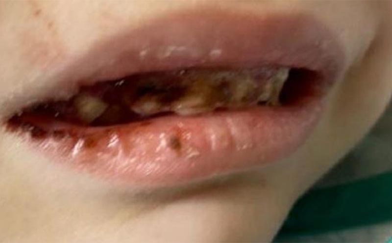
Exam was remarkable for a patient who was underweight and disheveled with bilateral knee swelling without overlying erythema, and a refusal to bear weight. Friable mucous membranes with bleeding and receding gingiva (Image 1) and scant scattered ecchymosis and bilateral petechiae with perifollicular hemorrhages were also noted.
Due to concern for neglect, a skeletal survey was completed. Due to the bilateral knee swelling, a point-of-care ultrasound was also obtained (Image 2).
Case Conclusion/Discussion
Radiographs of the distal femur and proximal tibia showed dense zones of provisional calcification (F: Frankel lines) with bony spurring noted at the bilateral distal femur and proximal tibia (P: Pelkan spurs) with underlying lucent metaphyseal bands (T: Trümmerfeld zone). Additionally noted were displaced fractures involving the distal metaphysis of the femur and tibia with overlying soft tissue swelling. Point of care ultrasound showed bilateral subperiosteal hemorrhage (arrow) and diaphyseal and metaphyseal fractures (arrowhead).
These radiographic findings, in conjunction with the clinical findings of mucosal bleeding, perifollicular petechiae, and arthralgias, suggested a diagnosis of scurvy.
Vitamin C (ascorbic acid) is an essential vitamin required for collagen synthesis. Scurvy is the disease caused by Vitamin C deficiency. Deficiency must persist for 8-12 weeks before symptoms develop.1,2 The diagnosis of scurvy can be made by history and physical exam findings confirmed by radiographs showing Frankel lines or Trümmerfeld zones, or a low serum ascorbic acid level.3 In our patient, their symptoms rapidly improved with ascorbic acid supplementation. As a result of their clinical presentation, imaging findings, and rapid clinical improvement with nutritional supplementation, the patient’s final diagnosis was that of scurvy.
References:
- Agarwal A, Shaharyar A, Kumar A, Bhat MS, and Mishra M. Scurvy in pediatric age group- A disease often forgotten? Journal of Clinical Orthopaedics and Trauma Jan 2015. (6) 101-107. http://dx.doi.org/10.1016/j.jcot.2014.12.003
- Smith A, Primio G. D., and Humphrey-Murto S. Scurvy in the developed world. CMAJ: Canadian Medical Association Journal, Aug 2011, 183(11), E752+. http://dx.doi.org/10.1503/cmaj.091938
- Ravi N, Gandhi N, Noel S, Chang K et al. Visual Diagnosis: Petechiae, Gingival Hyperplasia, Metaphyseal Lucencies, and Refusal to Ambulate in a 5-year-old Boy. Pediatrics in Review Dec 2020, 41 (12) e46-e49; http://dx.doi.org/10.1542/pit.2018-0080
Matthew Wiese, MD, PEM Fellow, PGY-6 a
Brendan Plotke, MS4 b
Puneet Gupta, MD a
Daniel Mirsch, MD ca Department of Pediatrics, Oishei Children’s Hospital, Buffalo NY.
b University at Buffalo Jacobs School of Medicine and Biomedical Sciences, Buffalo, NY.
c Department of Emergency Medicine, University at Buffalo Jacobs School of Medicine and Biomedical Sciences, Buffalo, NY.


